Congenital Extrahepatic Portosystemic Shunt
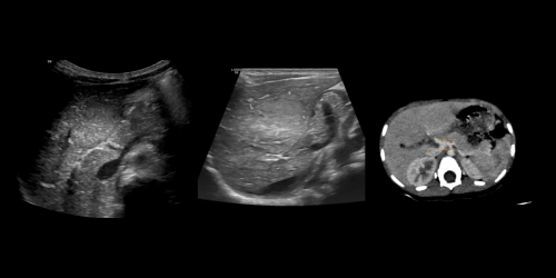
A 3-year-old male child with h/o of abdominal pain and USG shows hyperechoic area in liver.
Findings:
1. Portal vein: There is direct drainage of the non-branching portal vein into the retro hepatic IVC (both splenic vein and superior mesenteric vein forming acommon portal vein trunk draining into IVC) – Direct shunting blood fromthe portal venous system to the systemic venous system - Congenital portosystemicshunts (Extrahepatic)
2. An ill-defined hypodense lesion in left lobe of liver ( ultrasound revealed few (3-4) hyperechoic lesions in the liver) - nodular liver lesions, usually regenerative nodular hyperplasia
Diagnosis:
- Congenital extra-hepatic porto-systemic shunt (Type IB).
- Nodular liver lesions – likely regenerative nodular hyperplasia
Differentials:
For nodular liver lesion
- Focal nodular hyperplasia.
- Hepatocellular adenoma
Discussion:
Congenital extrahepatic portosystemic shunt (CEPS) is a condition in which the portomesenteric blood drains into a systemic vein, bypassing the liver through a complete or partial shunt.
In type 1 CEPS, there is complete diversion of portal blood into the systemic circulation (end-to-side shunt), with absent intrahepatic portal branches. Type 1 shunts are further classified into those in which the splenic vein (SV) and superior mesenteric vein (SMV) drain separately into a systemic vein (type 1a) and those in which the SV and SMV drain together after joining to form a common trunk (type 1b). In type 2 CEPS, the intrahepatic PV is intact, but some of the portal flow is diverted into a systemic vein through a side-to-side shunt
Nearly one-half of cases of CEPS are associated with nodular liver lesions, usually regenerative
nodular hyperplasia, focal nodular hyperplasia, or hepatocellular adenoma.
Details of Student:
Name:Dr. SANEESH PS
Email:[email protected]
Contact no. :7012072828
Institute/Hospital:ASTER MIMS ,Kannur
References:
- Alonso Gamarra, Eduardo & Parrón-Pajares, Manuel & Pérez, Ana & Prieto,Consuelo & Hierro, Loreto & López-Santamaría, Manuel. (2011). Clinical andRadiologic Manifestations of Congenital Extrahepatic Portosystemic Shunts: AComprehensive Review. Radiographics : a review publication of the Radiological Society of North America, Inc. 31. 707-22. 10.1148/rg.313105070.
- Gupta P, Sinha A, Sodhi KS, Lal A, Debi U, Thapa BR, Khandelwal N. Congenital Extrahepatic Portosystemic Shunts: Spectrum of Findings on Ultrasound,Computed Tomography, and Magnetic Resonance Imaging. Radiol Res Pract.2015;2015:181958. doi: 10.1155/2015/181958. Epub 2015 Dec 13. PMID:26858845; PMCID:PMC4691495.
More from AMI
Managing Adherent Placenta – Triple Doctor’s Story.
08/12/2022
Ectopia Cordis – A Case Study on a Rare Congenital Anomaly
29/11/2022
Call Fleming Syndrome
22/11/2022
Silk Vista Flow Diverter Stent – First time in Kerala @ Aster Medcity
22/11/2022
Celiac plexus block
20/12/2022
Mucormycosis
17/11/2023

AMI Expertise - When You Need It, Where You Need It.
Partner With Us
