


Teleradiology
Emerging Trends and Technologies in Medical Imaging
Introduction
The Inception of Medical Imaging
The accidental discovery of the X-ray in 1895 by Roentgen was the first major milestone in paving the path for modern-day medical imaging. It was considered a major milestone in aiding diagnosis as it allowed examining a patient's internal body parts without the need for any procedure for the very first time in the history of medical science. Roentgen was awarded the Noble Prize in Physics for the same. With time, human curiosity and the need to improvise diagnostic procedures led to the development of several sophisticated types of medical imaging procedures such as computed tomography (CT), magnetic resonance imaging (MRI), ultrasound (US), nuclear imaging, digital mammography and other hybrid scanners that allows three-dimensional imaging, four-dimensional imaging (three-dimensional imaging in real-time), functional imaging, and molecular imaging.
Medical imaging has now become quintessential in the modern healthcare scenario, its role dominating right from screening, aiding early diagnosis, treatment selection, and image-guided procedures to patient follow-up.
Breakthrough Advances in Medical Imaging
Advanced medical imaging technology has higher resolution, better reliability, is faster and exposes the patients to lower risk of radiation making it a safer choice to diagnose and manage complex medical conditions such as heart diseases, neurological conditions, cancers, etc.
Computed Tomography
CT is an advanced form of X-ray. Conventional CT involves passing X-ray radiation at different angles through the object of interest, which is then detected by an X-ray detector and processed by computers to recreate a series of 2D cross-sectional images.
CTs are one of the most widely used modalities for diagnosing infections, detecting conditions such as myocardial diseases, cerebral stroke, congenital heart diseases, idiopathic pulmonary fibrosis, dementia, etc and identifying cancers of the skeleton, liver, brain, stomach, lungs, etc. It is also useful in locating distant metastases, measuring changes in tumour masses before treatment and aiding procedures such as coronary artery bypass grafting.
Since its invention, many technological advances have been made in CT which have made it possible to scan much more efficiently, and faster within seconds and reduce the risk of radiation by more than half with improved image quality. The CT slices with modern machines are as thin as a millimetre.
Advanced bone medical imaging technologies include volumetric quantitative CT (vQCT), high-resolution CT (hrCT) and micro-CT (μCT). These advanced techniques not only provide 3D images of bone structures but also aids in measuring bone mineral density and bone strength, making them ideal for the management of osteoporosis and bone cancers.
Another advanced breast cancer imaging technology includes 3D USCT. Key advantages include a quick scan time of only 4 minutes of the entire breast volume and high image quality.
Magnetic Resonance Imaging
In MRI, a powerful magnet is used to generate a strong magnetic field around the patient's body and radiofrequency pulses are used to excite the photons inside the body. As the excited photons return to the resting stage, they generate signals that are captured and recreated as images.
Its superior soft tissue contrast and ability to create anatomical detail have made MRI a key diagnostic modality in oncology, where it is routinely used for staging and understanding the degree of metastasis. They are also ordered in diagnosing brain and spine infections, stroke, muscle degeneration, etc.
The whole-body MRI scan is commonly used for determining whole-body fat, soft tissue diseases, and polymyositis diseases and detecting pelvic, spine or femur lesions.
Advances in MRI include cardiac MRI and functional MRI. Cardiac MRI not only yields images of the heart morphology but also can assess cardiac function, making it a useful tool in establishing cardiomyopathy, congenital heart diseases, acute myocardial infarction, myocardial tumour and right ventricular dysplasia. Functional MRI is indicated in central nervous system diseases such as gliomas and in aiding surgeries by measuring brain activity.
Dynamic contrast-enhanced magnetic resonance (DCE-MRI) is an advanced technique for studying the tumour microenvironment. Multinuclear 3D solid-state MRI is another advanced technique that allows the recreation of tooth bone images and determining the composition and texture of the bones.
CT-MRI fusion imaging is yet another advanced tool used in cancer treatment, e.g. in neurosurgery, radiosurgery and hypofractionated radiotherapy. It is also used for monitoring brachytherapy in prostate and cervical cancer postimplant.
Ultrasound
The US procedure makes use of high-frequency sound waves above the range of human hearing to recreate organs and structures of the body in the form of images. The US can detect abnormalities in muscle functioning, thickness and fibre length, making it extremely suitable for diagnosing neuromuscular disorders.
The transvaginal US (TVS) is routinely used for monitoring and management of pregnancy and related complications. The US is also a simple, cheap and safe method for diagnosing complex renal masses.
Recent advances in US technology include 3D US, which generates 3D images and contrast-enhanced US (CEUS) which improves visualization of blood vessels and tissue characterization in solid organs. Ultrasound elastography can measure tissue stiffness and as tumour cells are stiffer than normal tissues, it can help in tumour detection and characterization in the breast, liver and thyroid gland tissues.
High-intensity focused US (HIFU) and MR-guided focused US (MRGFUS) are used to focally heat and ablate target tissues, without damaging the surrounding normal tissues. These methods are used for conditions such as medication-resistant Parkinson’s disease, uterine fibroids, prostate cancer, etc.
High-resolution US which is also known as ultrasound biomicroscopy (UBM) uses high-frequency sound waves above 35 MHz to provide high-resolution images of the human eye, making it a good choice of diagnostic for ocular trauma, glaucoma, eye lens displacement, hyphemia, cataract, etc. Low-frequency US systems are being used to aid non-invasive drug delivery through the human skin, such as insulin, erythropoietin, and interferon-gamma molecules.
Positron emission tomography
The PET scan involves the intravenous administration of a radioactive tracer in the patient, that emits low-energy photons. These low-energy photons are detected by the PET camera which is ultimately converted to 3D images. The key use of PET scans is to study biological processes inside the body, such as cell proliferation and DNA replication. Heart diseases, Alzheimer's disease, cancer, and Huntington's disease can be diagnosed using the PET scan.
The PET scan also measures the concentration of various metabolites such as amino acids, sugar, fatty acids, etc inside the cell and is being used as a diagnostic tool to diagnose atherosclerosis, cancer, schizophrenia, etc.
A combination of PET-CT is often used in the management of cancer, inflammatory and infectious diseases.
Single-Photon Emission Computed Tomography
Single-photon emission computed tomography (SPECT) makes use of radioactive tracers, which emits gamma rays to generate 3D images of various body parts with high accuracy. The advanced SPECT technology with its improved features can detect deep and very small fractures in the patient. It can be also used for imaging myocardial disorders, bone metabolism, and neurochemical brain imaging. SPECT/CT is a powerful tool in diagnosing neuropsychiatric diseases, bone tumours, bone trauma and bone infections.
Digital mammography
Mammography involves the use of low-energy X-rays to screen and diagnoses breast cancer. Conventional mammography has been replaced by digital mammography, which is faster and more accurate in aiding diagnosis in younger patients less than 50 years of age, with denser breasts or with breast implants and in premenopausal women. The key advantages of digital mammography over conventional techniques include that it can be stored electronically for future reference and sent easily for a second opinion. The images can be manipulated for better clarity and visualization and it is associated with a lower average radiation dose.
Current Trends in Medical Imaging Technology
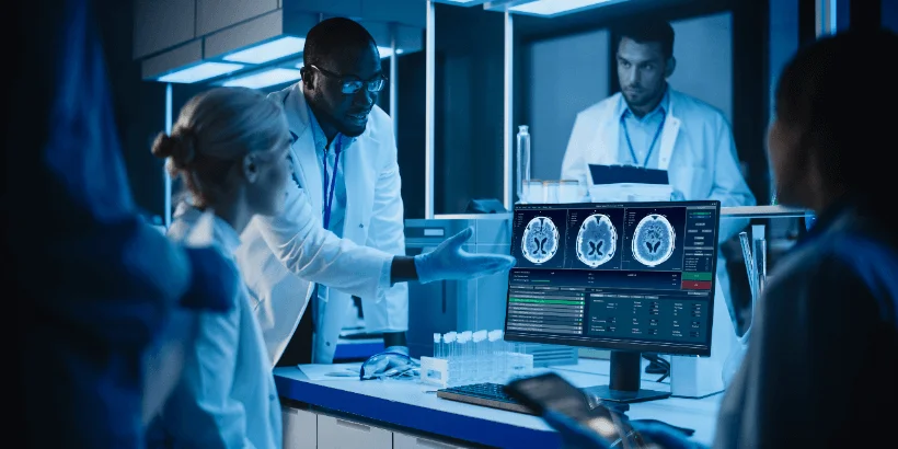
The advent of technology has transformed healthcare and made a significant impact in improving patient safety and aiding diagnosis and treatment delivery. Real-time patient information with just a click of a finger for faster and more efficient management, reduced mortality and hospital admission rates. The primary drivers of advanced imaging modalities include artificial intelligence (AI), machine learning, the Internet of medical technology and deep learning.
The integration of AI in imaging procedures such as in advanced CT scans, MRIs, and USs has helped to carry out repetitive tasks with almost 99% accuracy and minimal error in a more cost-effective manner as compared to conventional modalities. Healthcare practitioners are preferring AI-based solutions for calculating risks and designing treatment plans based on individual case profiles to deliver the most effective therapy at minimal cost to the patient.
The integration of AI in mobile apps and watches has led to the collection of massive amounts of medical data including heart rate, sleep pattern, activity rate, etc which can help patients to predict their risk of developing certain chronic medical conditions. Wearables ECG has demonstrated the potential to identify patients at risk and reduce the rates of emergency visits.
Teleradiology is another current trend in medical radiology, which makes use of telecommunication systems to transmit radiological images from anywhere in the globe to a specialist radiologist at another place for appropriate interpretation of imaging studies, such as X-rays, CT scans, MRI, etc. Considering the rising demand for imaging in establishing an accurate diagnosis, especially in critical care settings and the global shortage of speciality radiologists, teleradiology holds the potential to bridge this gap and improve patient care significantly.
Conclusion
With the advancement of newer technology, the future of medical imaging is limitless. With time, more advanced technology will allow even faster, safer and more efficient imaging with a more advanced way of storing, sharing and interpreting medical data.
Aster Medical Imaging, a leading teleradiology company, adopt the proven and latest technologies clubbed with globally accepted safety protocols. We are well equipped with the latest medical equipment, modern technology and infrastructure and host the best of brains in the field of expert radiology. We deliver outstanding value to hospitals and medical centres seeking to optimize their resources with extensive onsite imaging services. More than 100 of our medical imaging units work to deliver the highest quality of patient care to our partners.
Our Joint Commission (JC) accredited NABH solutions eliminate the challenge of setting up your own radiology departments while working closely with you to effect the best patient outcomes. Our personalized fits allow you the flexibility, and coverage to choose the services that best suit your patient needs. Our Interventional Radiology offerings are customized to cater to vastly differing needs, meeting your current, and future demands with ease.
We are well equipped and excel in the field of neuroimaging, onco-imaging, interventional radiology, musculoskeletal imaging, cardiac radiology, paediatric radiology, emergency radiology, breast imaging & mammography, etc. Our specialist radiologists work in close association with consulting and referring physicians of the patient, ensuring optimal outcomes. They are actively involved in the diagnosis and treatment process with all stakeholders in the patient management plan.
More from AMI
Cardiovascular Imaging: Expanding Access to Advanced Heart Care with Teleradiology
14/04/2026
Why Hospitals Prefer Teleradiology Outsourcing for Accurate and Timely MSK Injury Reports
14/11/2025
MRI vs Ultrasound Imaging for Sports Injuries: Which Test Is Right?
24/02/2026
How to Choose a Prospective Teleradiology Service Provider
07/08/2023
Emerging Techniques in Radiology By Dr. Namita
10/11/2022
What Is Teleradiology? Improving Access, Availability, and Affordability of Radiology in India
22/01/2026
Sports-Related Injuries and the Importance of Radiology
30/01/2023
Behind The Scenes of Teleradiology: How Digital Imaging Is Changing Diagnostic Medicine
16/10/2023
CT MRI Brain scans and Their Role in Neurological Diagnosis
26/12/2025
MRI imaging for Memory Loss: Understanding Alzheimer’s and Dementia
26/12/2025
Why Accurate Cancer Report Images Are Critical for Treatment Planning
25/03/2026
How Teleradiology Supports Musculoskeletal Pain Specialists in Diagnosis
20/08/2025
How Teleradiology Reporting Services Are Revolutionizing Frozen Shoulder Diagnosis and Reporting
17/11/2025
What is Diagnostic Radiology? Understanding its Tests and Procedures
14/08/2023
Teleradiology's Contribution to Timely Emergency Diagnoses
04/10/2023
Imaging of Ventricular Septal Defect (VSD): Role of Cardiac Imaging in Detection & Surgical Planning
21/01/2026
Atherosclerosis Imaging: How Advanced Cardiovascular Imaging Enables Early Heart Disease Detection
21/01/2026
Revolutionizing Indian Healthcare: Unlocking the Potential of Teleradiology in Remote Areas
27/09/2023
MRI Spine Thoracic: How Teleradiology Enhances Faster, Accurate Diagnosis
29/09/2025
Radiology Practices
30/11/2022
Intra- Operative 3D Imaging With O- Arm Making Complex Spine Surgeries Safe and Accurate
30/11/2022
Imaging In Pregnancy
18/01/2023
Early Detection of Stress Fractures Through Advanced Musculoskeletal Service Imaging
24/02/2026
Advances In Neuroradiology
06/01/2023
Paediatric Traumatic Brain Injury (TBI) Imaging: CT vs MRI Brain Imaging in Detecting Subtle Brain Injuries in Children
26/12/2025
The Future of MRI: Advancements and AI Integration in Online MRI Reporting
19/05/2025
Scope of Radiologist in India
29/08/2023
Empowering Radiologists-Teleradiology Redefines the Role of Imaging Specialist
09/10/2023
How Fast Reporting (TAT) Saves Lives in Oncology Images
25/03/2026
Imaging Instrumentation Trends In Clinical Oncology
26/06/2023
Coronary Plaque Formation: How Advanced Cardiology Imaging Identifies High-Risk Plaques
30/01/2026
Radiology Online Reporting: Transforming Diagnosis with Speed and Accuracy
31/07/2025
10 Strategies To Prevent Burnout In Radiology
09/01/2023
Unlocking the Knee: How Tele-Reporting Service Empowers Clinicians
25/11/2025
How Teleradiology Solutions Help Hospitals Cut Turnaround Time
20/08/2025
Improvement of Patient Care Through Teleradiology
03/07/2023
How Aster Medical Imaging’s Nighthawk Service Enhances Brain Neuroimaging
29/09/2025
PET-CT: The Gold Standard in Cancer Detection – What Patients Must Know
27/03/2026
How to Increase the Efficiency of the Radiology Equipment
21/08/2023

AMI Expertise - When You Need It, Where You Need It.
Partner With Us