Thoracic Radiology Services
The credentialed team of radiologists at AMI provide screening and accurate diagnosis with state-of-the-art imaging modalities, advanced technology and expertise.
Thoracic imaging radiologists specialize in the diagnosis and treatment of chest and lung diseases. The top-notch thoracic subspecialists at AMI are recognized both nationally and internationally, and are qualified with advanced fellowships in cardiothoracic imaging.
Their specialties include:
- Pulmonary infections
- Primary and secondary thoracic malignancies
- Infectious diseases – Tuberculosis and COVID-19 virus
- Mediastinal masses
- Pleural disease
- Emphysema
- Interstitial lung disease
- Lymphoma
- Occupational disorders
- Diseases like asthma, bronchiectasis, and tracheomalacia, which affect the large and small airways
Our wide range of chest-related diagnostic examinations include:
- Chest radiography
- Chest fluoroscopy
- Computed tomography (CT)
- High-resolution CT
- Low-dose screening CT
- Magnetic Resonance Imaging (MRI)
Our thoracic radiologists work closely with other physicians like:
- Primary care physicians
- Surgeons
- Pulmonologists
- Gastroenterologists
- Urologists
- Gynecologists
- Radiation and Medical Oncologists

What do we offer at AMI?
How Thoracic Radiology Reporting Can Improve Your Throughput Using Our Services
Quality
Reporting standards followed as per guidelines from the American College of Radiology (ACR) & The Royal College of Radiologists (RCR)
On-Time Reports
Reliable, and accurate reports with less turn-around time. 99% of the emergency reports are delivered in less than 1 hour.
24/7 Compliance
Internationally certified radiologists with Sub-specialty expertise are available 24×7 for 365 days a year.
FAQs
A thoracic radiologist specializes in interpreting medical images related to the thoracic region, which includes the chest and its internal structures. These radiologists play a crucial role in diagnosing and managing a wide range of conditions affecting the lungs, heart, mediastinum, and chest wall. Their expertise encompasses the interpretation of various imaging modalities, such as chest X-rays, computed tomography (CT) scans, magnetic resonance imaging (MRI), and nuclear medicine studies. Thoracic radiologists are skilled in identifying abnormalities such as pulmonary nodules, masses, infections, and congenital anomalies. They play a key role in the evaluation of cardiovascular diseases, including the assessment of coronary arteries and cardiac chambers. Additionally, they contribute to the diagnosis and staging of thoracic cancers and assist in guiding interventional procedures like lung biopsies or drainage of pleural effusions using imaging guidance. Collaboration with pulmonologists, cardiologists, oncologists, and other healthcare professionals is common for thoracic radiologists, ensuring comprehensive patient care in the context of thoracic diseases. Their specialized knowledge and interpretation skills are essential in providing accurate diagnoses and contributing to effective treatment plans for patients with thoracic conditions.
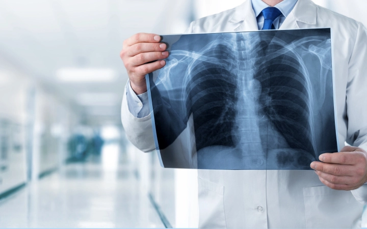
A thoracic radiograph, commonly known as a chest X-ray, is a diagnostic imaging technique that provides a two-dimensional view of the chest and its internal structures. This non-invasive and widely used imaging modality involves exposing the chest to a small amount of ionizing radiation to create an image capturing the anatomy of the lungs, heart, ribs, and surrounding structures. Thoracic radiographs are invaluable for detecting and evaluating a variety of medical conditions, including lung infections, pneumonia, tuberculosis, pulmonary masses, fractures, and heart-related abnormalities. The images obtained from a thoracic radiograph aid healthcare professionals, including thoracic radiologists, pulmonologists, and other clinicians, in making accurate diagnoses and determining appropriate treatment plans. Thoracic radiographs are often the initial step in the diagnostic process, providing a quick and accessible assessment of the chest to guide further evaluation or intervention if necessary.
Recent developments in thoracic radiology have been marked by significant strides in technology, improving diagnostic accuracy and patient care. Artificial intelligence (AI) applications have gained prominence, with machine learning algorithms assisting radiologists in the interpretation of chest imaging, enhancing efficiency, and aiding in the early detection of thoracic abnormalities. Advanced imaging techniques, such as dual-energy CT and high-resolution CT, continue to evolve, allowing for more detailed visualization of pulmonary structures and improved characterization of lung diseases. Functional imaging modalities, including positron emission tomography (PET) combined with CT, provide valuable insights into both the structural and metabolic aspects of thoracic conditions, aiding in the diagnosis and staging of lung cancer and other diseases. Lung cancer screening programs, utilizing low-dose CT scans, have become more widespread, contributing to the early detection of malignancies and improved outcomes. Furthermore, a personalized medicine approach in thoracic oncology is emerging, leveraging molecular imaging and genetic profiling to tailor treatments based on the unique characteristics of individual tumors. These advancements collectively underscore the ongoing efforts to enhance diagnostic capabilities, refine treatment strategies, and ultimately improve patient outcomes in the field of thoracic radiology.

AMI Expertise - When You Need It, Where You Need It.
Partner With Us

